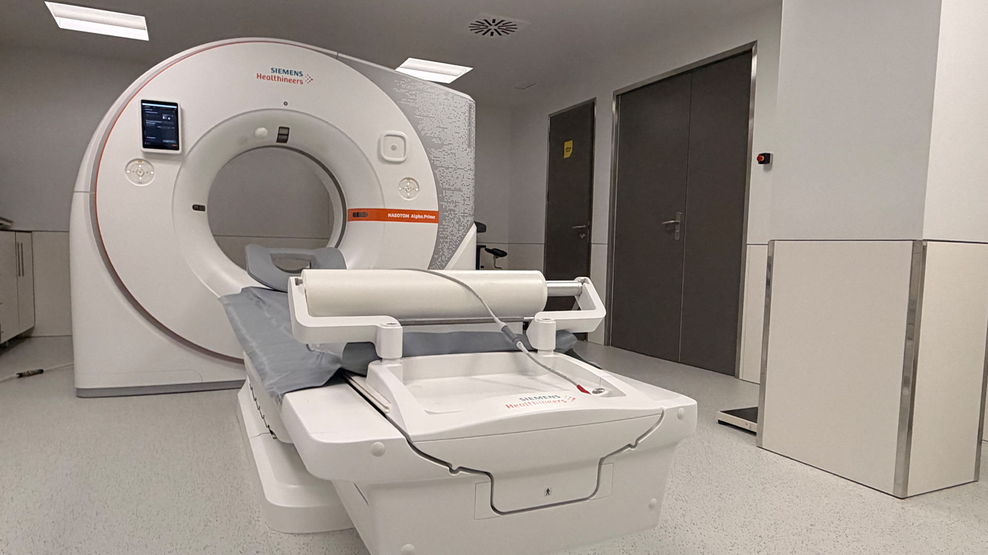
Lisbon, 3 August 2026 – Portugal now has the first Photon Counting technology in operation in the country: a new generation of Computed Tomography, considered the greatest technological development in this field in recent decades. By strengthening diagnostic capacity and supporting more precise clinical decisions, this new technology may also help reduce the need for additional complementary examinations and invasive procedures across multiple areas of Medicine.
Unlike conventional Computed Tomography, Photon Counting technology measures each X-ray photon individually, instead of integrating all the energy received by the detector. This technological difference makes it possible to obtain more anatomically detailed images, reduce artefacts that make examinations harder to interpret, and improve the characterisation of lesions, tissues and anatomical structures, including small structures and even in particularly demanding situations, such as patients with extensive arterial calcifications or coronary stents.
For specialists, this development represents more than an improvement in image quality. By providing better information from the first examination, it may contribute to more accurate diagnoses and, in certain clinical contexts, reduce the need for additional studies, some of them invasive.
The technology also makes it possible to reduce radiation and/or contrast doses in certain clinical protocols, while maintaining high levels of diagnostic quality — a benefit that is particularly relevant for patients who require periodic examinations or who are more vulnerable to cumulative exposure to radiation or contrast agents.
“We are facing a highly significant development in diagnostic imaging. It is not simply about obtaining better-quality images. We are able to obtain more and better diagnostic information, observe anatomical structures with a much higher level of detail and increase confidence in the diagnosis and, consequently, in decision-making,” says Luís Curvo Semedo, Radiologist at Joaquim Chaves Saúde.
In the cardiovascular field, one of the main advantages lies in the assessment of coronary artery disease, particularly in patients with significant calcifications or stents, situations in which the conventional technique may have limitations.
“In this field, one of the main added values of this new technology is the reduction of artefacts caused by calcifications and stents, allowing a more accurate assessment of the inside of the coronary arteries. It is possible to evaluate and clarify clinical situations that previously could involve some diagnostic uncertainty or in which CT angiography examinations were even non-diagnostic,” notes Carla Saraiva, Radiologist dedicated to cardiac imaging at Joaquim Chaves Saúde.
In the respiratory field, this technology may also represent an important advance in the characterisation of very early pulmonary changes. Its high spatial resolution allows a more detailed assessment of small pulmonary nodules and early-stage changes, particularly in the context of screening or suspected lung cancer — a capability that may contribute to earlier and more precise diagnoses in situations where image detail truly makes a difference. In the assessment of interstitial lung diseases, this technology may also enable timelier diagnoses by detecting changes in very small anatomical structures at an earlier stage.
Luis Lebre, Clinical Director of Joaquim Chaves Saúde, adds that “another advantage is the reduction of the cumulative risk associated with repeated examinations, which is especially important for patients who require periodic monitoring (e.g. lung cancer screening, oncological disease follow-up), for children and young people, who are more sensitive to the long-term effects of radiation, and for pregnant women, when the examination is strictly necessary, as well as the possible reduction of oncological risk due to a lower accumulated dose”.
For Luís Curvo Semedo, the impact of this technology is transversal across different medical specialties: “more than a technological development, we are facing a new ability to obtain diagnostic information from imaging. This allows different pathologies to be better characterised, supports more informed therapeutic decisions and improves coordination between medical specialties, for the benefit of the patient.”
The benefits of Photon Counting technology extend to multiple areas of medicine, including Cardiology, Pulmonology, Oncology, Neurology and Orthopaedics, among many other specialties in which diagnostic precision plays a decisive role.
The technology is installed at Joaquim Chaves Saúde’s Miraflores Clinic, where the first NAEOTOM Alpha Prime system from Siemens Healthineers is now available and in operation in Portugal.
In addition to private activity and services provided under agreements with different insurers, the examinations are also available to users of the National Health Service, under a regional agreement covering all Local Health Units in the Lisbon and Tagus Valley region, upon referral issued by one of these units.